Adenomyomatosis Gallbladder ICD-10, Symptoms, Causes, Treatment
Gall bladder adenomyomatosis is a common benign condition, with features such as tumors of unknown origin characterized by hyperplastic wall changes, and often termed as hyperplastic cholecystosis. Adenomyomatosis of the gall bladder and the associated Rokitansky Aschoff sinuses may include the gall bladder as a focal, segmental or diffuse form.
A barrier or annular thickening causes peripheral narrowing of the lumen often associated with adenomyomatosis and must be distinguished from congenital folds of the wall of the gallbladder which is usually thinner and smoother and is located at the bottom while adenomyomatosis may include a part of the gallbladder. Adenomyomatosis is most common in middle aged women and its aetiology remains unclear.
Thickening of the wall of the gallbladder may suggest the diagnosis of gallbladder cancer, resulting in unnecessary treatment or even surgery. Gallbladder compartmentalization in the hourglass type of adenomyomatosis often makes difficult to identify the distal compartment during radiographic scanning or contribute to incomplete cholecystectomy when only the distal half of the gallbladder is removed at surgery.
Symptoms of adenomyomatosis are so obvious however some commonly observed symptoms are bloating, biliary colic, vague abdominal pain, intolerance to fatty foods and dyspepsia. In a small amount of patients adenomyomatosis associated with fever and jaundice.
Adenomvomatosis (or diverticulitis of gall bladder) is an acquired hyperplastic condition characterized by excessive epithelial surface proliferation with extensive invaginations or diverticles (so called sinusoidal Rokitansky Aschoff) protruding into the muscular layer of the wall of gall bladder. Adenomyomatosis causes the gallbladder to thicken or diffuse the wall containing small cyst type spacesat cross sectional imaging. These cysts as spaces can result in a "pearl necklace" sign in T2 weighted MRI. The state has a propensity for the bottom of the gallbladder. The central gallbladder can also be affected, as a result of typical "hourglass" configuration.
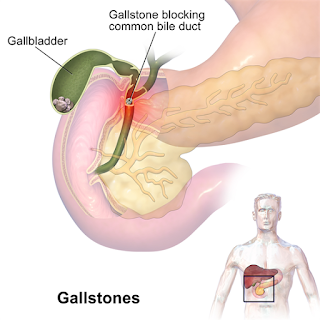
Adenomyomatosis itself remains asymptomatic unless it is associated with other pathological condition like gallstones or cholecystectomy. If such conditions are associated with adenomyomatosis then surgery is mainstay to resolve underlying cause. However, pain might be used in case of severe pain. Adenomyomatosis itself is serious medical issue but it must be followed up for further diagnosis to find out main problem.
Following code is used for Adenomyomatosis in ICD-10
K82.8--Other specified diseases of gallbladder--billable
A barrier or annular thickening causes peripheral narrowing of the lumen often associated with adenomyomatosis and must be distinguished from congenital folds of the wall of the gallbladder which is usually thinner and smoother and is located at the bottom while adenomyomatosis may include a part of the gallbladder. Adenomyomatosis is most common in middle aged women and its aetiology remains unclear.
Thickening of the wall of the gallbladder may suggest the diagnosis of gallbladder cancer, resulting in unnecessary treatment or even surgery. Gallbladder compartmentalization in the hourglass type of adenomyomatosis often makes difficult to identify the distal compartment during radiographic scanning or contribute to incomplete cholecystectomy when only the distal half of the gallbladder is removed at surgery.
Adenomyomatosis Symptoms
Symptoms of adenomyomatosis are so obvious however some commonly observed symptoms are bloating, biliary colic, vague abdominal pain, intolerance to fatty foods and dyspepsia. In a small amount of patients adenomyomatosis associated with fever and jaundice.
Adenomyomatosis Causes
Adenomvomatosis (or diverticulitis of gall bladder) is an acquired hyperplastic condition characterized by excessive epithelial surface proliferation with extensive invaginations or diverticles (so called sinusoidal Rokitansky Aschoff) protruding into the muscular layer of the wall of gall bladder. Adenomyomatosis causes the gallbladder to thicken or diffuse the wall containing small cyst type spacesat cross sectional imaging. These cysts as spaces can result in a "pearl necklace" sign in T2 weighted MRI. The state has a propensity for the bottom of the gallbladder. The central gallbladder can also be affected, as a result of typical "hourglass" configuration.
Adenomyomatosis Treatment
Adenomyomatosis ICD-10
Following code is used for Adenomyomatosis in ICD-10
K82.8--Other specified diseases of gallbladder--billable
Adenomyomatosis Gallbladder ICD-10, Symptoms, Causes, Treatment
 Reviewed by Simon Albert
on
December 23, 2019
Rating:
 Reviewed by Simon Albert
on
December 23, 2019
Rating:


