Chorionic Villi Definition, Development, Miscarriage, Histology
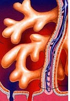
Chorionic villi are the small outgrowths which looks like finger present inside the placenta. Chorionic villi also contains genetic material which is same as present inside the cells of fetus. The term chorion means a membrane present during pregnancy between mother and fetus. And this membrane contains villi on it. This membrane is responsible for the covering of embryo in many mammals, birds and reptiles. Chorion also helps in the development of fetus. Chorionic villi are considered as the main functional unit of placenta. It also responsible for the separation of blood of fetus from mother within placenta.
During the development of fetus, there are many connections between mother and fetus and chorionic villi is one of these connections. It is formed by combined tissues from mother and fetus. Chorionic villi are microscopic structures rich in capillaries looks like finger. It is the barrier which transfer nutrients from mother to the fetus. Blood supplied to the chorion through the arteries and deoxygenated blood gets out through veins. These villi are the channel for the nutrients from mother and channel for transferring waste from fetus. The length of villi is long enough to enter the endometrium.

At the initial stage of development of villi, each villi follow the same procedure. Later, the structural morphology and the functions associated are changed. In week two (primary villi), trophoblastic cells form villi. In week three (secondary villi), the mesoderm from extra embryonic turns into villi and covers the whole sac also forms chorionic plate. In week four(tertiary villi), capillary network forms in villi which connects with the vessels of placenta and forms a stalk which connects both. The stalk contains:
Chorionic villi miscarriage occurs when the villi are immature. There is a very little risk of miscarriage along with pregnancy every time. CVS sampling during pregnancy can be the cause of miscarriage. CVS test is normal in pregnancy but there are also little chances of miscarriage. The ratio of miscarriage to CVS is 3:200. The chances of miscarriage are also seen after amniocentesis. But the chances of miscarriage in CVS are high as compare to amniocentesis, the reason behind this is CVS sampling is done at the initial stage of pregnancy. Any kind of suspected problem may also lead to miscarriage.
Chorionic villi are rich in capillaries and connective tissues that connects the fetus to blood of mother. The cells involved in the connective tissues are known as fibroblasts. Some macrophages are also present in the chorionic villi and these macrophages are known as hofbauer cells. Chorionic villi contains dual blood supply. All the villi have same basic structure. Villi are covered with an epithelial layer known as syncytiotrophoblast. Villous cytotrophoblast are located between upper syncytiotrophoblast and the base membrane. Chorionic villi are the base of stem cells. The baseline separates villous stroma from the trophoblastic membrane.
During the development of fetus, there are many connections between mother and fetus and chorionic villi is one of these connections. It is formed by combined tissues from mother and fetus. Chorionic villi are microscopic structures rich in capillaries looks like finger. It is the barrier which transfer nutrients from mother to the fetus. Blood supplied to the chorion through the arteries and deoxygenated blood gets out through veins. These villi are the channel for the nutrients from mother and channel for transferring waste from fetus. The length of villi is long enough to enter the endometrium.

Chorionic Villi Development:
At the initial stage of development of villi, each villi follow the same procedure. Later, the structural morphology and the functions associated are changed. In week two (primary villi), trophoblastic cells form villi. In week three (secondary villi), the mesoderm from extra embryonic turns into villi and covers the whole sac also forms chorionic plate. In week four(tertiary villi), capillary network forms in villi which connects with the vessels of placenta and forms a stalk which connects both. The stalk contains:
- Stem villi
- Terminal villi
- Branched villi
Chorionic Villi Miscarriage
Chorionic villi miscarriage occurs when the villi are immature. There is a very little risk of miscarriage along with pregnancy every time. CVS sampling during pregnancy can be the cause of miscarriage. CVS test is normal in pregnancy but there are also little chances of miscarriage. The ratio of miscarriage to CVS is 3:200. The chances of miscarriage are also seen after amniocentesis. But the chances of miscarriage in CVS are high as compare to amniocentesis, the reason behind this is CVS sampling is done at the initial stage of pregnancy. Any kind of suspected problem may also lead to miscarriage.
Chorionic Villi Histology
Chorionic villi are rich in capillaries and connective tissues that connects the fetus to blood of mother. The cells involved in the connective tissues are known as fibroblasts. Some macrophages are also present in the chorionic villi and these macrophages are known as hofbauer cells. Chorionic villi contains dual blood supply. All the villi have same basic structure. Villi are covered with an epithelial layer known as syncytiotrophoblast. Villous cytotrophoblast are located between upper syncytiotrophoblast and the base membrane. Chorionic villi are the base of stem cells. The baseline separates villous stroma from the trophoblastic membrane.
Chorionic Villi Definition, Development, Miscarriage, Histology
 Reviewed by Simon Albert
on
March 24, 2017
Rating:
 Reviewed by Simon Albert
on
March 24, 2017
Rating:


